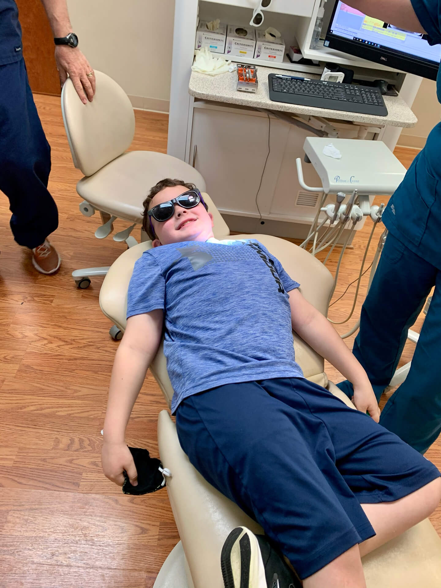
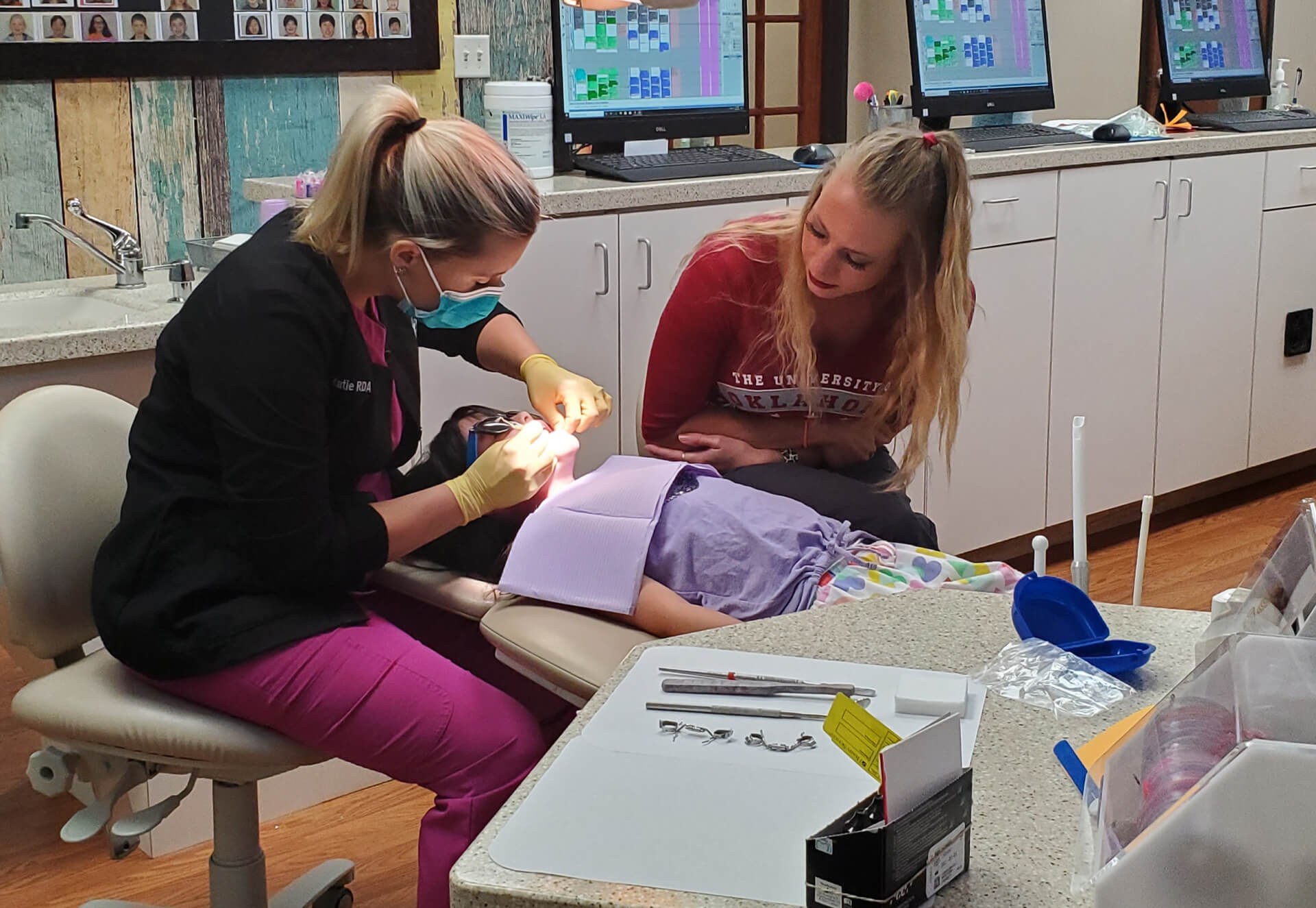
Laser Dentistry


Our system is one of the most advanced laser dental treatments available. The laser tip allows a dental surgeon to direct a concentrated beam of light at a target tissue and produce precise results in an exceptionally gentle manner. Using a surgical laser over a blade has many advantages:
- Faster healing time and less trauma to teeth and gums.
- Less anesthetic for most procedures.
- Treating dental needs in more than one part of the mouth in one visit.
- Making dental care a more relaxing experience for our patients.
Tongue Tie / Ankyloglossia
Approximately 16% of infants are born with an anatomical defect known as Ankyloglossia, or tongue-tie. It results from a shortened frenulum. This is the strip of mucous membrane attaching the underside of the tongue to the bottom of the mouth.
This anatomic abnormality can cause a host of problems for a developing child. The tongue is an important morphofunctional organ which is largely responsible for proper orofacial development and growth. The restriction ankyloglossia produces has been linked to an abnormal suction swallow / chewing pattern in infants and newborns and frequently leads to speech disorders. A short lingual frenum also results in low tongue position and tongue thrusting which contributes to the formation of high / narrow palatal vault (one of the features favoring collapse of the upper airway during sleep). This breathing dysfunction can result in malocclusion, prognathism, and distortion of the “harmonic face”. This will cause the mid third of the face to appear smaller than the upper and lower thirds. Tongue tie can also result in mouth breathing, which may be a factor in tonsillar enlargement. This increase in upper airway resistance causes soft tissue inflammation and adenoid / tonsillar growth. Hypertrophic adenotonsils, in turn, can obstruct the airflow and exacerbate mouth breathing, and contribute to the abnormal orofacial growth. This is how being tongue-tie can create a vicious circle ultimately causing sleep disordered breathing (SDB) and obstructive sleep apnea (OSA). A breathing dysfunction can also lead to a host of other developmental issues for a young child.
Tongue Tie / Ankyloglossia
As the permanent teeth begin to emerge from the gum line, pediatric dentists may recommend that a labial frenectomy be performed for orthodontic, periodontal, or aesthetic reasons. The labial frenum is the soft tissue that attaches the upper or lower lip to the mouth. When a thick or abnormally attached frenulum is present, a gap between the teeth can form. If left untreated, this gap, also called a diastema, can become permanent, creating spacing issues for other teeth as well as gum recession. Plaque accumulation can also increase and result in chronic gingivitis and periodontal pockets. Bone loss is a possible long- term consequence of failing to correct this early on in life.
Gingival Hypertrophy
This is the dental term for swollen or overgrown gum tissue. This condition can hurt and cause a patient’s teeth to appear abnormally short or uneven. Hypertrophy can occur for a variety of reasons. In most cases, pediatric patients experience this phenomenon from irritation associated with poor oral hygiene. Pediatric patients undergoing orthodontic treatment can develop this condition if they don’t keep their braces clean. A majority of this overgrowth improves with proper brushing habits and or the removal of braces. If the tissue doesn’t shrink back enough after the source of irritation is removed, we surgically remove it with the laser tip.
Treatment (Frenectomy / Gingivectomy)
There are many options for treatment depending on the patient’s age and thickness of the frenum / hypertrophic gingiva. Outdated methods involve excising or snipping the tissue with a scalpel or sharp scissors after a local anesthetic is administered. For the frenectomy procedure, dissolvable stitches are used to close the surgical site.
The newer and more advanced surgical technique of frenum and overgrown gingival excision involves the use of a laser. The laser is far less invasive than the scalpel resulting in minimal bleeding and trauma to the mucosa. Wounds heal faster and patients experience less post-operative pain. Stitches are also not required making this a far superior technique.
Laser Pulpotomy
A vital pulpotomy is a single-stage procedure in which a pediatric dentist surgically removes the infected portion of the baby tooth nerve. This infection results from bacteria in a deep cavity “eating” its way through the outer tooth and coming into contact with the sterile “inner tooth nerve”. If allowed to develop, bacteria will infect an entire tooth rendering it non-restorable. Eventually this “cavity bacteria” will tunnel out of the tooth, through the jaw, and infect the face. At this point the infection becomes life threatening and patients require I.V. antibiotics and hospitalization to resolve the infection. The baby tooth will then have to be removed prematurely resulting in abnormal jaw and adult tooth development. The pulpotomy is therefore done as a means of eliminating tooth decay and preserving the vitality and function of the remaining tooth nerve. Caustic chemicals such as formaldehyde are typically used to disinfect the infected pulp once the cavity was removed. The laser option eliminates the need for this dated approach by vaporizing only infected pulp tissue and leaving behind healthy pulp. The baby tooth is then capped with a “tooth hat”, commonly referred to as a crown, which eventually falls out with the baby tooth when an adult tooth is ready to replace it. It’s as if the cavity never happened.
We love hearing from our amazing patients.

